Abstract
The involution of the postpartum bovine uterus is accompanied by bacterial invasion. Studies show that most, if not all, uterine cavities are bacterially contaminated in the immediate post-partum period. The culture at this time will usually produce a wide range of bacteria, including Actinomyces pyogenes, Streptococcus spp., Staphylococcus spp. and Clostridium spp., Coliforms and Gram-negative anaerobes.
The research is part of a larger study that aimed to isolate and identify potentially pathogenic bacteria from uterine secretions and their role in postpartum infections. To isolate and identify the uterine flora, swabs (n=160) were collected from the lumen of 32 dairy cattle, between 3 to >21 DIM (days in milk). Bacterial microflora has been monitored for 5 weeks. The samples were passed through all stages of the microbiological examination and the results revealed the presence of the species Streptococcus agalactiae and Enterococcus faecalis.
At the first examination Streptococcus agalactiae and Enterococcus faecalis were isolated in 8 (25%) of 32 samples, and Streptococcus agalactiae in monoculture was isolated in 4 (12, 5%) of 32 samples. At the second and third examination the number of Streptococcus agalactiae in mono-culture decreased. At 21 days after parturition Streptococcus agalactiae and Enterococcus faecal-is were isolated in 11 (34, 38%) of 32 samples and Streptococcus agalactiae in monoculture was found in 9 (28,12%) of cows. The presence of these bacterial species with pathogenic potential for cattle and humans, highlights a possible zoonotic risk.
Keywords: Dairy cows; Group B Streptococcus; Enterococcus faecalis, Uterine infection
Introduction
After parturition in cows, there are a number of changes in the uterus that are accompanied by bacterial con-tamination. The abnormal development of the uterine involution process determines the bacterial multiplication, and results in uterine infections [15, 31]. Members of the genus Bacillus, Streptococcus, and Enterococcus, in addition to coagulase-negative Staphylococci, are among the most frequently isolated intrauterine bacteria and have been described as potential or opportunistic pathogens [1, 5, 12, 25, 27, 28].
Streptococcus agalactiae represents group B Streptococcus (GBS) and is a common colonizer of the genital and gastrointestinal tracts [9]. Until 1930, GBS was considered an important microbial agent of bovine origin, which was responsible for the appearance of mastitis [3]. Its pathogenicity, but especially its zoonotic role was highlighted in other species of mammals, reptiles and fish [4, 26]. Over time, GBS has become an important pathogen for human pathology and screenings have been performed to highlight the degree of human contamination [10, 18].
Group B Streptococcus is a major cause of neonatal infections, including pneumonia and meningitis in new-borns and young infants less than three months. After birth, in women Streptococcus agalactiae can be the cause of endometritis with blood dissemination and secondary endocardial or meningeal localization. In immunodeficiency adults with diseases like diabetes or cancer Streptococcus agalactiae can cause infections of the skin, soft tissue, os-teomyelitis, pneumonia or septicemia [6].
Some studies have described that interspecies transmission of Streptococcus agalactiae between cattle and humans is possible [13]. The presence of these organisms in the first weeks after calving should not be considered abnormal. In a healthy postpartum uterus, the number of bacteria should decrease rapidly, and bacteria will be absent or present only in very small numbers within three to four weeks of farrowing [21].
Enterococci are commensal of the human and animals GI tract. In humans is also associated with urinary tract infections, surgical wound infections, endocarditis and sepsis [30].
Enterococcus faecalis and Enterococcus faecium are responsible for the majority of enterococcal infections. The treatment of enterococcal infections imposes difficulties due to their ability to acquire resistance to many antibiotics, especially vancomycin. The difficulties of treatment in enterococcus infections come from the fact that antibiotics are used in raising cattle, which leads to the transmission of resistant bacteria from animals to humans through the food chain [2].
Materials and Methods
The aim of this study was to monitor the bacterial flora for 5 weeks postpartum by microbiological examination.The study was conducted on 2 dairy farms in county Iasi. The predominant breed was Black and White Ro-manian cows and Holstein was also present. The number of studied cows was 32 (16 per farm). All cows were examined between 3 to 45 DIM by vaginoscopy and bacteriological examinations.
After vaginoscopy, samples were collected with a cotton swab on a metal rod (40 cm length) for bacteriological examination. The samples were inoculated into Mueller Hinton Broth (Oxoid) and incubated at 37oC for 24h aerobically. The isolation and direct identification of group B Streptococcus was carried out on StrepBSelect Agar (chromogenic agar plate), Columbia CNA +5% Sheep Blood (Bio-Rad Laboratories). The plates were incubated for 24-48 hours at 37 o C under aerobic conditions.
According to the manufacturer, on StrepB Select Agar the specific colonies of GBS are blue, and the purple colonies indicate Enterococcus spp. Each type of colony was transplanted into Muller Hinton Agar (Bio Rad La-boratories). The strains of Enterococcus spp. have been transplanted onto Bile Esculin Agar (BEA) which is a dif-ferential and selective medium and is mainly used to discern group D Streptococci and Enterococci based on the organism’s potential to hydrolyze esculin. To differentiate Enterococcus faecalis and Enterococcus faecium, their ability to utilize pyruvate was tested. After incubation at 37°C for 24 hours, the streptococcal strains were sero-logically tested. The Prolex™ Strep (Pro-Lab Diagnostis) was used to confirm the serogroup.
Results
Gynecological examination
At the examination of the cows on day 21 post-partum, 17/32 were diagnosed with clinical endometritis, and at more than 45 days all animals showed clinical signs of endometritis. Based on the character of vaginal discharge more than half of the animals had abnormal vaginal mucus.
Isolation and identification of bacteria
A total of 160 samples were included in the analysis. The isolation and direct identification of Streptococcus agalactiae and Enterococcus spp. was carried out on chromogenic agar plate, StrepBSelectTMAgar (Bio-Rad Labora-tories) (Figure 1).
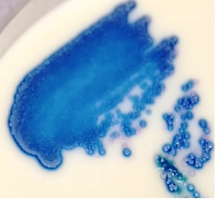
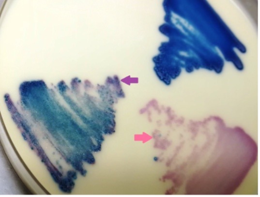
Figure 1. (Left) Streptococcus agalactiae (blue colonies) on StrepBSelect Agar
(Right) Enterococcus spp. (purple arrow) and Lactobacillus (pink arrow) growing on StrepBSelect Agar
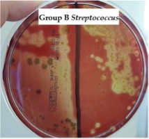
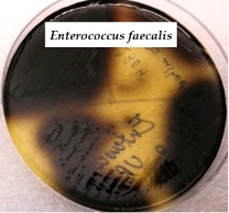
To confirm the presence of Streptococcus agalactiae blue colonies from StrepBSelect Agar (Bio-Rad Laboratories). were inoculated on blood agar to see beta-hemolytic activity (Figure 2). The confirmation for purple colonies from StrepBSelect Agar was been made on a selective medium for enterococci, BEA (Bile Esculine Azide Agar)(Bio-Rad Laboratories). This media contains bile to suppress the growth of gram pozitive and negative bacteria. The positive reaction is confirmed by the brown color around the colonies (Figure 3).
|
|
Figure 2. Streptococcus agalactiae, beta-hemolytic, Columbia CNA +5% Sheep Blood |
Figure 3. Colonies of Enterococcus faecalis on Bile Esculin Azide Agar(BEA) |
It has also, the serological identification of streptococcal strains was carried out using commercial latex particle agglutination (Prolex Streptococcal Grouping Kit )(Figure 4).
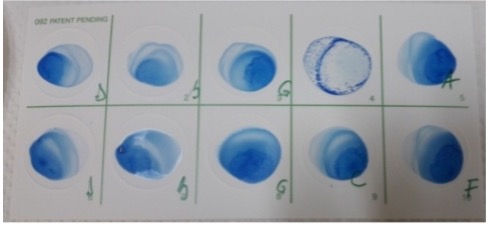
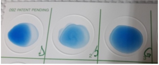
Figure 4. Group confirmatory serology Streptococcus agalactiae ( gr.B), Enterococcus faecalis ( gr.D)
Bacterial isolates from the same cows during the five examination were compared. The prevalence of bacteria is indicated in Table 1; at the first examination Streptococcus agalactiae and Enterococcus faecalis were isolated in 8 (25%) of 32 samples, and Streptococcus agalactiae in monoculture was isolated in 4 (12,5%) of 32 samples. At the second and third examination the number of Streptococcus agalactiae in monoculture decreased.
At 21 days after parturition Streptococcus agalactiae and Enterococcus faecalis were isolated in 11 (34,38%) of 32 samples and Streptococcus agalactiae in monoculture was found in 9 (28,12%) of 32 cows.
Cows with an intrauterine infection of Streptococcus agalactiae and Enterococcus faecalis at first examination were at higher risk to be re-diagnosed with the same bacterial species at next four examination. Streptococcus agalactiae in monoculture was continually isolated in 1 cow during the five examination. A new infection with Streptococcus agalactiae in monoculture occurred in 8 cows at 21 day after parturition (Table 1).
Table 1. Incidence of the species Streptococcus agalactiae and Enterococcus faecalis isolated from uterine discharges
|
Tested animals |
32 |
32 |
32 |
32 |
32 |
|
3 day |
9 day |
15 day |
21 day |
>21 day |
|
|
Streptococcus agalactiae + Enterococcus faecalis |
25% |
18,8% |
9,37% |
34,37% |
25% |
|
Enterococcus faecalis |
3,12% |
0% |
0% |
6,25% |
3,12% |
|
Streptococcus agalactiae |
12,5% |
6,25% |
3,12% |
28,12% |
6,25% |
Discussion
The objective of the present study was to illustrates the growth density of bacterial content in the uterus of dairy cows between 3 to >21 DIM and not just isolation.
Bacterial examination of the vagina and cervix has been evaluated based on abnormal vaginal discharge.
Animals that received any systemic antibiotic treatment between or after parturition were not included in the study to avoid false-negative bacteriological findings.
The clinical examination of the genital tract included visual inspection of leaks, transrectal palpation, followed by vaginoscopy examination. Before vaginal examination, the vulva was cleaned with dry paper. An autoclaved Polansky vaginal speculum with 3 blades was moistened with vaseline and inserted into the vagina. The visual examination was support by a flashlight.
Following the gynecological examination at 21 days postpartum, 53.13% of the animals were diagnosed with clinical endometritis. Other studies reported a prevalence of clinical endometritis between 10% and 20% [16, 23, 24]; however, there are other studies with a prevalence > 40% [17,19, 27].
Uterine bacterial contamination decreases logarithmically with increasing postpartum days [7, 8, 22]. In this study this observation could be confirmed for Streptococcus agalactiae and Enterococcus faecalis but at 21 days after partu-rition a significant increase was found in the case of this isolated bacterial species.
Enterococcus faecalis was isolated from infected postpartum uterus in cows and had been described as potential or opportunistic pathogens, finding supported by other studies [11, 29].
The isolation of Streptococcus agalactiae in the first three weeks after parturition was 12,5%, 6,25%, 3,12%, results that highlight a decrease in contamination. At 21 DIM Streptococcus agalactiae was isolated in monoculture in 28,12% of cows.
The results of this study describe the possibility of contamination of the uterine cavity by caregivers or veterinarians who perform treatments at this level. The way in which pathogens get in the uterus is unclear, whether they come directly from the skin of the cow, the caregiver's/veterinarian hands or from the environment [20], through exposure to excreta from humans.
Given the location of Streptococcus agalactiae of the human throat, gut and urogenital tract, the most plausible route of transmission from cattle to humans is through exposure to feces, with or without use of gloves [16].
This cycle of contamination and recontamination by direct contact between humans and cows indicates that bacterial strains may favor certain routes of contamination and it is justified that control and treatment measures for Streptococcus agalactiae in dairy herds may fail [14].
As a recommendation for limiting GBS infection and colonization, compounds with antimicrobial or inhibitory activity, obtained from plants or of synthetic origin, could be administered. These agents could create a uterine barrier and not destroy the commensal flora at the same time [16].
Conclusion
All bacterial isolates used in the present study come from both farms. This indicate that subsequent research using specific methods for identifying and patterning species and subspecies are needed to elucidate the impact of Streptococcus agalactiae on uterine health.
Group B Streptococcus is a pathogen with major involvement in neonatal infections in humans and is carried asymptomatically by a large part of the population but is also recognized as a pathogen of dairy cattle in parts of Europe.
Due to the small number of samples investigated, further studies on a larger number of isolates are needed to document the colonization with Group B Streptococcus of the genital tract in Romanian dairy cows, given that no similar information is known.
The isolation and identification of this bacterial species in 28,12% of the cows at 21 days after parturition can be considered an alarm signal in the context of the suspicion of interspecies transmission from cattle to humans.
Enterococcus faecalis is recognized for its ability to acquire antibiotic resistance and its presence in proportion of 6,25 % of animals from the study may be responsible for therapeutic failures in cows with puerperal disease.
Refferences
[1] Ballas, P., Reinländer, U., Schlegl, R., Ehling-Schulz, M., Drillich, M., & Wagener, K. Characterization of intrauterine cultivable aerobic microbiota at the time of insemination in dairy cows with and without mild endometritis. Theriogenology 2021, 159, 28-34.
[2] Beukers, A. G., Zaheer, R., Goji, N., Amoako, K. K., Chaves, A. V., Ward, M. P., & McAllister, T. A. Comparative genomics of Enterococcus spp. isolated from bovine feces. BMC microbiology 2017, 17(1), 1-18.
[3] Bisharat, N., Crook, D. W., Leigh, J., Harding, R. M., Ward, P. N., Coffey, T. J. Jones, N. Hyperinvasive neonatal group B streptococcus has arisen from a bovine ancestor. Journal of Clinical Microbiology 2004, 42(5), 2161-2167.
[4] Brochet, M., Couvé, E., Zouine, M., Vallaeys, T., Rusniok, C., Lamy, M. C. Glaser, P. Genomic diversity and evolution within the species Streptococcus agalactiae. Microbes and Infection 2006, 8(5), 1227-1243.
[5] Carneiro, L. C., Cronin, J. G., & Sheldon, I. M. Mechanisms linking bacterial infections of the bovine endometrium to disease and infertility. Reproductive biology 2016, 16(1), 1-7.
[6] Cobo-Angel C.G., Jaramillo-Jaramillo A.S., Palacio-Aguilera M., Jurado-Vargas L., Calvo-Villegas E.A., Ospina-Loaiza D.A., Rodriguez-Lecompte J.C., Sanchez J., Zadoks R., Ceballos-Marquez A. Potential group B Streptococcus interspecies transmission between cattle and people in Colombian dairy farms. Sci Rep. 2019, Oct 1; 9(1):14025.
[7] Drugociu, D. and Drugociu D.S. Patologie genitală şi a glandei mamare la animale. Editura" Ion Ionescu de la Brad 2015.
[8] Elliott, L., McMahon, K. J., Gier, H. T., & Marion, G. B. (1968). Uterus of the cow after parturition: bacterial content.American Journal of Veterinary Research 1968, 29(1), 77-81.
[9]] Hanna, M., & Noor, A. Streptococcus Group B. StatPearls 2020 [Internet].
[10] Ho, M., Chang, Y. Y., Chang, W. C., Lin, H. C., Wang, M. H., Lin, W. C., & Chiu, T. H. Oral Lactobacillus rhamnosus GR-1 and Lactobacillus reuteri RC-14 to reduce Group B Streptococcus colonization in pregnant women: a randomized controlled trial.Taiwanese Journal of Obstetrics and Gynecology 2016, 55(4), 515-518.
[11] Hou, R., Chen, C., Fu, Y., Liu, Y., Li, L., Hao, Y., & Huang, S. Analysis of microbial flora in dairy cows vagina, and the isolation and identification of Lactobacillus.Zhongguo Weishengtaxixue Zazhi/Chinese Journal of Microecology 2011, 23(5), 393-397.
[12] Huszenicza, G., Fodor, M., Gacs, M., Kulcsar, M., Dohmen, M. J. W., Vamos, M., Szita, G. Uterine bacteriology, resumption of cyclic ovarian activity and fertility in postpartum cows kept in large‐scale dairy herds. Reproduction in Domestic Animals 1999 , 34(3‐4), 237-245.
[13] Jensen, N. E. Experimental bovine group-B streptococcal mastitis induced by strains of human and bovine origin. Nordisk veterinaermedicin 1982 34.12: 441-45
[15] Knudsen, L. R. V., Karstrup, C. C., Pedersen, H. G., Angen, Ø., Agerholm, J. S., Rasmussen, E. L., Klitgaard, K. An investigation of the microbiota in uterine flush samples and endometrial biopsies from dairy cows during the first 7 weeks postpartum. Theriogenology 2016, 86(2), 642-650.
[16] LeBlanc, S. J., Duffield, T. F., Leslie, K. E., Bateman, K. G., Keefe, G. P., Walton, J. S., & Johnson, W. H. Defining and diagnosing postpartum clinical endometritis and its impact on reproductive performance in dairy cows. Journal of dairy science 2002, 85(9), 2223-2236.
[17] Knudsen, L. R. V., Karstrup, C. C., Pedersen, H. G., Angen, Ø., Agerholm, J. S., Rasmussen, E. L., Klitgaard, K. An investigation of the microbiota in uterine flush samples and endometrial biopsies from dairy cows during the first 7 weeks postpartum. Theriogenology 2016, 86(2), 642-650.
[18] Patras, K. A., Nizet, V. Group B streptococcal maternal colonization and neonatal disease: molecular mechanisms and preventative approaches. Frontiers in Pediatrics 2018, 6, 27.
[19] Pleticha, S., Drillich, M., & Heuwieser, W. Evaluation of the Metricheck device and the gloved hand for the diagnosis of clinical endometritis in dairy cows. Journal of dairy science 2009, 92(11), 5429-5435.
[20] Prunner I., Wagener K., Pothmann H., Ehling-Schulz M., Drillich M. Risk factors for uterine diseases on small- and medium-sized dairy farms determined by clinical, bacteriological, and cytological examinations. Theriogenology 2014; 82:857–65
[21] Pulfer, K. W., and R. L. Riese. "Treatment of postpartum metritis in dairy cows." Iowa State University Veterinarian 1991, 53.1: 6.
[22] Runceanu, L., Cotea, C., Drugociu, D., Roşca, P. Reproducţie, obstetrică şi ginecologie veterinară.Ed.” Ion Ionescu de la Brad Iaşi. 2001
[23] Sheldon, I. M., & Dobson, H. Postpartum uterine health in cattle. Animal reproduction science 2004, 82, 295-306.
[24] Sheldon, I. M., Lewis, G. S., LeBlanc, S., Gilbert, R. O. Defining postpartum uterine disease in cattle. Theriogenology 2006, 65(8), 1516-1530.
[25] Sikra, A. A., Rîmbu C., and Drugociu D. Impact of staphylococcus aureus and methicillin-resistant staphylococcus aureus (mrsa) on uterine disease in dairy cattle after parturition.Scientific papers veterinary medicine 2020 , 63(3), 261-267
[26] Sun, J., Fang, W., Ke, B., He, D., Liang, Y., Ning, D., Deng, X. Inapparent Streptococcus agalactiae infection in adult/commercial tilapia. Scientific reports 2016, 6(1), 1-11.
[27] Wagener, K., Grunert, T., Prunner, I., Ehling-Schulz, M., Drillich, M. Dynamics of uterine infections with Escherichia coli, Streptococcus uberis and Trueperella pyogenes in post-partum dairy cows and their association with clinical endometritis. The Veterinary Journal 2014, 202(3), 527-532.
[28] Wang, Y., Ametaj, B. N., Ambrose, D. J., & Gänzle, M. G. Characterisation of the bacterial microbiota of the vagina of dairy cows and isolation of pediocin-producing Pediococcus acidilactici. BMC microbiology 2013, 13(1), 1-11.
[29] Wang, J., Sun, C., Liu, C., Yang, Y., & Lu, W. Comparison of vaginal microbial community structure in healthy and endometritis dairy cows by PCR-DGGE and real-time PCR. Anaerobe 2016, 38, 1-6.
[30] Willems, R. J., Top, J., Marga van Santen, D., Coque, T. M., Baquero, F., Grundmann, H., & Bonten, M. J. Global spread of vancomycin-resistant Enterococcus faecium from distinct nosocomial genetic complex. Emerging infectious diseases 2005, 11(6), 821.
[31]Williams, E. J., Fischer, D. P., Pfeiffer, D. U., England, G. C., Noakes, D. E., Dobson, H., & Sheldon, I. M. Clinical evaluation of postpartum vaginal mucus reflects uterine bacterial infection and the immune response in cattle. Theriogenology 2005, 63(1), 102-117.